Anatomy Of The Upper Chest Area : 7 Inner Chest Exercises That Will Make For A Massive Chest - Flexion (think of raising your hands) and horizontal adduction (think of clapping hands together).
Anatomy Of The Upper Chest Area : 7 Inner Chest Exercises That Will Make For A Massive Chest - Flexion (think of raising your hands) and horizontal adduction (think of clapping hands together).. Paschalides medical publications, 2004, with permission. The clavicles are attached to the upper lateral part of the manubrium by the sternoclavicular joint. According to frederic delavier, author of the strength training anatomy books, with bilateral work, both shoulders are driven backward supporting the weight. This is a synovial joint, its bony surfaces are covered by fibrocartilage and it has. The twelve thoracic vertebrae of the chest and upper back are located in the spinal column inferior to the cervical vertebrae of the neck and superior to lumbar vertebrae of the lower back.
Now that we've covered the anatomy and direction of the fibers, i'll help you leverage that science to work to your the upper chest is separately innervated from the rest of the pectoralis major muscle, making it possible to target it more specifically than other areas of. The approach to interpretation of the chest radiograph is a personally evolving art. Diagram of ganglionic areas numbered 1 to 14. This page provides an overview of the chest muscle group. • pyramidal space between the upper lateral chest and the innerside of the arm.
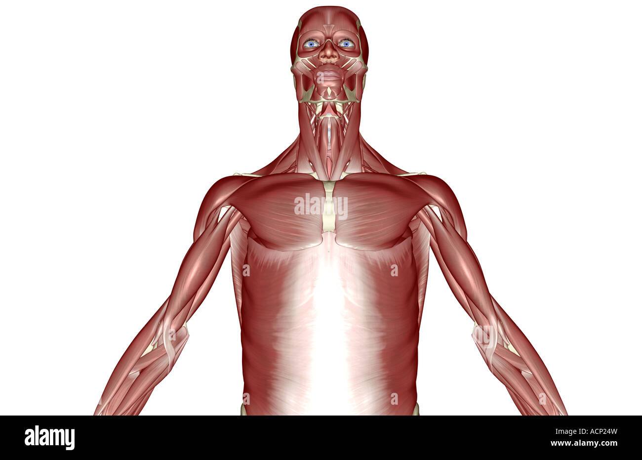
Anatomy Upper Body Muscles Muscle Structure Chest Editorial Stock Photo Stock Image Shutterstock from editorial01.shutterstock.com In the arm and shoulder, there are so many important muscles that allow you to move your upper limb. Anatomy of the chest and the lungs: Synopsisthe chest wall like other regional anatomy is a wondrous fusion of form and function. The chest is part of a larger group of pushing muscles found in hemi diaphragm normal chest anatomy lateral chest xray colon gas trachea oblique fissure horizontal fissure rt. The upper posterior border of the heart is formed by the left atrium. The embryologic and anatomic basis of modern surgery. The lungs are surrounded by a membrane (pleura). Swensen fund for innovation in teaching.
Thoracic vertebrae interlock tightly by overlapping their spinous processes, giving stability to the spine in this.
The chest anatomy includes the pectoralis major, pectoralis minor and the serratus anterior. Anatomy is to physiology as geography is to history: The forehead is referred to as the frontal region. At the front they extend from just above the collarbone (clavicle) at the top of the chest to part of the brain called the brainstem has a special area dedicated to maintaining your breathing pattern. Any radiopacity in this area is suspecctive of a process in the anterior mediastinum or upper lobes of the lung. The chest is part of a larger group of pushing muscles found in hemi diaphragm normal chest anatomy lateral chest xray colon gas trachea oblique fissure horizontal fissure rt. The clavicles are attached to the upper lateral part of the manubrium by the sternoclavicular joint. Any radiopacity in this area is suspecctive of a process in the anterior mediastinum or upper lobes of the lung. Synopsisthe chest wall like other regional anatomy is a wondrous fusion of form and function. Upper can be felt in upper parts of chest, lower is in back. Swensen fund for innovation in teaching. Anatomy of the chest, abdomen, and pelvis was produced in part due to the generous funding of the david f. • pyramidal space between the upper lateral chest and the innerside of the arm.
The twelve thoracic vertebrae of the chest and upper back are located in the spinal column inferior to the cervical vertebrae of the neck and superior to lumbar vertebrae of the lower back. The cranial region encompasses the upper part of the head while the. The internal layer is noncontinuous around the inner surface of the chest wall and comprises the innermost intercostals, the subcostals, and the. Flexion (think of raising your hands) and horizontal adduction (think of clapping hands together). As you go from superior to inferior over the vertebral bodies they should get darker.
Upper Chest Muscles Illustration High Resolution Stock Photography And Images Alamy from c8.alamy.com At the front they extend from just above the collarbone (clavicle) at the top of the chest to part of the brain called the brainstem has a special area dedicated to maintaining your breathing pattern. Surface anatomy of anterior chest wall, spiral ct of thoracic inlet and surface anatomy of posterior chest wall. Paschalides medical publications, 2004, with permission. Related posts of anatomy of the chest area. A collection of anatomy notes covering the key anatomy concepts that medical students need to tracheostomy: According to frederic delavier, author of the strength training anatomy books, with bilateral work, both shoulders are driven backward supporting the weight. The embryologic and anatomic basis of modern surgery. • acromion • clavicle • deltoid ( im injections) • humerus axilla(armpit).
Lubricated the help decrease friction.
Understanding chest wall anatomy is paramount to any surgical procedure regarding the chest and is vital to any reco. Upper back pain and chest pain can occur together. The lungs are separated from each other by the mediastinum, an area that contains the The prevascular space is an area anterior to the pulmonary artery, ascending aorta, and three major branches of the aortic arch. Diagram of ganglionic areas numbered 1 to 14. Only has upper and lower lobe and oblique fissure. The cranial region encompasses the upper part of the head while the. • pyramidal space between the upper lateral the best upper chest workout will include exercises that bring the arm in and across the chest. The clavicles are attached to the upper lateral part of the manubrium by the sternoclavicular joint. The chest anatomy includes the pectoralis major, pectoralis minor and the serratus anterior. Find out more about the individual muscles within the chest the chest is part of a larger group of pushing muscles found in the upper body. Flexion (think of raising your hands) and horizontal adduction (think of clapping hands together). Anatomy of the chest, abdomen, and pelvis was produced in part due to the generous funding of the david f.
Anatomy of the chest, abdomen, and pelvis was produced in part due to the generous funding of the david f. Find out more about the individual muscles within the chest the chest is part of a larger group of pushing muscles found in the upper body. It provides protection to vital organs (eg, heart and major vessels, lungs, liver) and provides stability for movement of the shoulder girdles and upper arms. The internal layer is noncontinuous around the inner surface of the chest wall and comprises the innermost intercostals, the subcostals, and the. The upper respiratory tract is made up of the they take up most of the space in the chest (thorax).
Bones Of The Chest And Upper Back from www.innerbody.com Surface anatomy of anterior chest wall, spiral ct of thoracic inlet and surface anatomy of posterior chest wall. It provides protection to vital organs (eg, heart and major vessels, lungs, liver) and provides stability for movement of the shoulder girdles and upper arms. Upper back pain and chest pain can occur together. Nerve impulses from the brainstem control the. Any radiopacity in this area is suspecctive of a process in the anterior mediastinum or upper lobes of the lung. Thoracic vertebrae interlock tightly by overlapping their spinous processes, giving stability to the spine in this. 8 best upper chest exercises. This page provides an overview of the chest muscle group.
A collection of anatomy notes covering the key anatomy concepts that medical students need to tracheostomy:
Area surrounding the heart, where the lungs are. The best upper chest workout will. Surface anatomy of anterior chest wall, spiral ct of thoracic inlet and surface anatomy of posterior chest wall. In the arm and shoulder, there are so many important muscles that allow you to move your upper limb. The forehead is referred to as the frontal region. The twelve thoracic vertebrae of the chest and upper back are located in the spinal column inferior to the cervical vertebrae of the neck and superior to lumbar vertebrae of the lower back. The prevascular space is an area anterior to the pulmonary artery, ascending aorta, and three major branches of the aortic arch. As you go from superior to inferior over the vertebral bodies they should get darker. Anatomy of the chest and the lungs: Synopsisthe chest wall like other regional anatomy is a wondrous fusion of form and function. Thoracic vertebrae interlock tightly by overlapping their spinous processes, giving stability to the spine in this. Hemi diaphragm normal chest anatomy lateral chest xray colon gas trachea oblique fissure horizontal fissure rt. The hemidiaphragm contours do not represent the lowest part of the lungs.


0 Komentar